Yttrium »
PDB 1dde-3ph5 »
3n9d »
Yttrium in PDB 3n9d: Monoclinic Structure of P. Aeruginosa Ligd Phosphoesterase Domain
Protein crystallography data
The structure of Monoclinic Structure of P. Aeruginosa Ligd Phosphoesterase Domain, PDB code: 3n9d
was solved by
S.Shuman,
P.Nair,
P.Smith,
with X-Ray Crystallography technique. A brief refinement statistics is given in the table below:
| Resolution Low / High (Å) | 28.29 / 2.30 |
| Space group | C 1 2 1 |
| Cell size a, b, c (Å), α, β, γ (°) | 90.997, 56.585, 43.503, 90.00, 118.33, 90.00 |
| R / Rfree (%) | 21.2 / 26.3 |
Other elements in 3n9d:
The structure of Monoclinic Structure of P. Aeruginosa Ligd Phosphoesterase Domain also contains other interesting chemical elements:
| Manganese | (Mn) | 1 atom |
Yttrium Binding Sites:
The binding sites of Yttrium atom in the Monoclinic Structure of P. Aeruginosa Ligd Phosphoesterase Domain
(pdb code 3n9d). This binding sites where shown within
5.0 Angstroms radius around Yttrium atom.
In total 2 binding sites of Yttrium where determined in the Monoclinic Structure of P. Aeruginosa Ligd Phosphoesterase Domain, PDB code: 3n9d:
Jump to Yttrium binding site number: 1; 2;
In total 2 binding sites of Yttrium where determined in the Monoclinic Structure of P. Aeruginosa Ligd Phosphoesterase Domain, PDB code: 3n9d:
Jump to Yttrium binding site number: 1; 2;
Yttrium binding site 1 out of 2 in 3n9d
Go back to
Yttrium binding site 1 out
of 2 in the Monoclinic Structure of P. Aeruginosa Ligd Phosphoesterase Domain
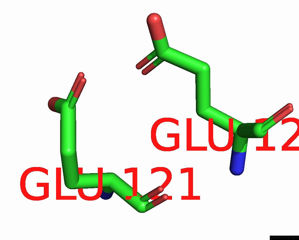
Mono view
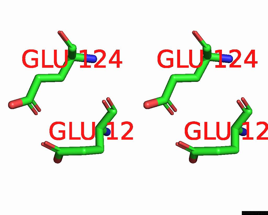
Stereo pair view
Mono view
Stereo pair view
A full contact list of Yttrium with other atoms in the Y binding
site number 1 of Monoclinic Structure of P. Aeruginosa Ligd Phosphoesterase Domain within 5.0Å range:
|
Yttrium binding site 2 out of 2 in 3n9d
Go back to
Yttrium binding site 2 out
of 2 in the Monoclinic Structure of P. Aeruginosa Ligd Phosphoesterase Domain
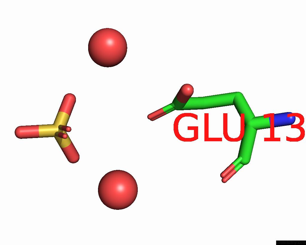
Mono view
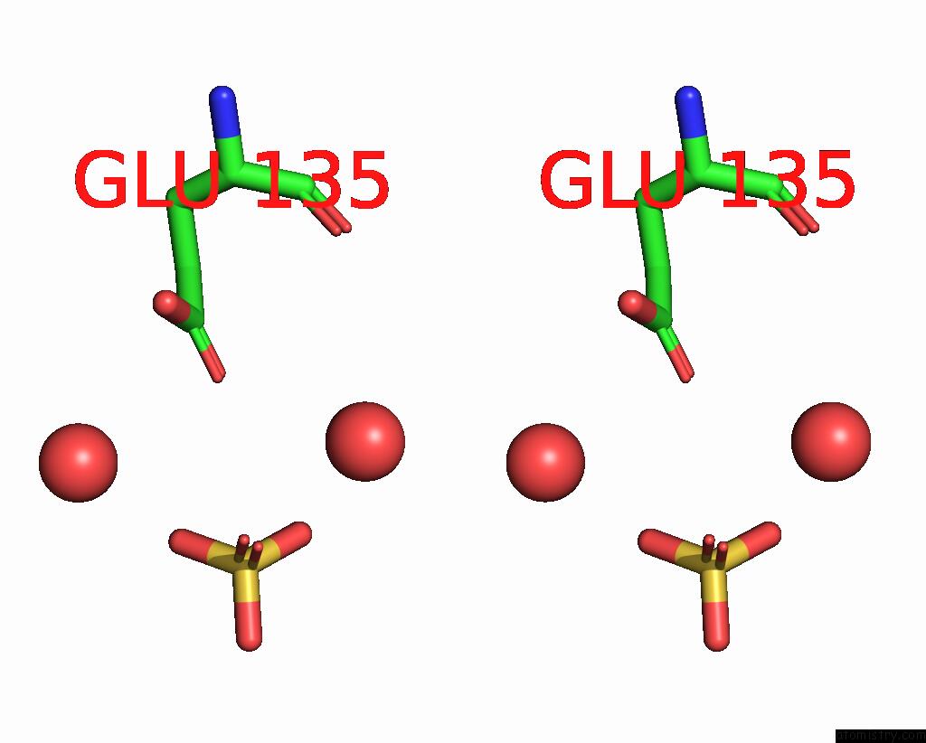
Stereo pair view
Mono view
Stereo pair view
A full contact list of Yttrium with other atoms in the Y binding
site number 2 of Monoclinic Structure of P. Aeruginosa Ligd Phosphoesterase Domain within 5.0Å range:
|
Reference:
P.A.Nair,
P.Smith,
S.Shuman.
Structure of Bacterial Ligd 3'-Phosphoesterase Unveils A Dna Repair Superfamily Proc.Natl.Acad.Sci.Usa V. 107 12822 2010.
ISSN: ISSN 0027-8424
PubMed: 20616014
DOI: 10.1073/PNAS.1005830107
Page generated: Sat Oct 12 20:35:41 2024
ISSN: ISSN 0027-8424
PubMed: 20616014
DOI: 10.1073/PNAS.1005830107
Last articles
Cl in 3UXDCl in 3UY9
Cl in 3UZC
Cl in 3UXH
Cl in 3UXE
Cl in 3UX0
Cl in 3UWT
Cl in 3UUW
Cl in 3UWD
Cl in 3UVC